Interpreting PET/CT Images to Offer the Best Cancer Treatment
February 17, 2017
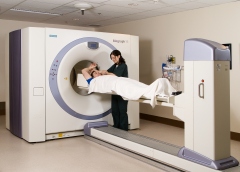
Positron Emission Tomography/Computed Tomography, or PET/CT, is an imaging method that uses a sophisticated machine to generate images of patients following the injection of very small amounts of a medical isotope.
As a Radiologist and Nuclear Medicine physician in the Functional Imaging Department at BC Cancer, my primary role is to interpret the PET/CT images obtained on one of the two scanners at the Vancouver Centre and then generate a report for the referring physician. In addition to looking at the images produced on the day, I review all the relevant previous imaging as well as laboratory and clinical information on each patient in order to provide the most comprehensive interpretation possible.
What I do in addition to reporting varies on a day-to-day basis, from prioritizing requisitions, to reviewing cases with my clinical colleagues to participating in local and provincial multidisciplinary tumour conferences. Every day is a little bit different, which keeps things interesting!
The Functional Imaging Department is involved in over 20 active clinical research trials, some of which are internal, such as the NaF trial comparing sodium fluoride bone scans to standard 99m Tc bone scans in patients with prostate cancer. The majority, however, are clinical trials where imaging plays a part, all with the goal of increasing our knowledge to better treat patients with cancer.
Pete

State-of-the-Art Scanning Technology a First for Canada
Support BC Cancer – Vancouver’s Technology Transformation — a suite of best-in-class diagnostic, imaging and radiation equipment, including the Next Gen PET/CT.
Donate Today